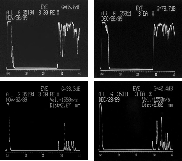
Case # 1 Case # 2 Case # 3
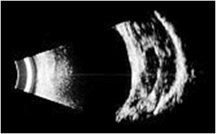
B-scan:
diagnoses
UBM- B-scan: diagnoses
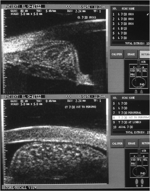
Iris Cyst
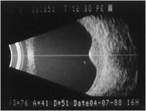
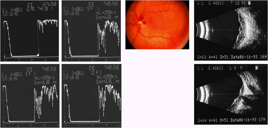
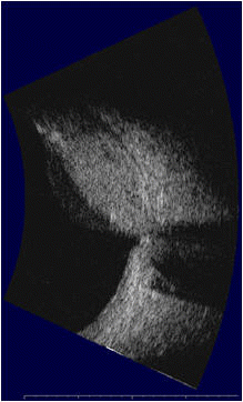
B-scan: shows Choroidal Tumor
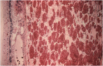
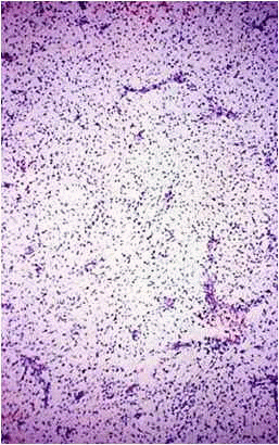
Choroidal Melanoma
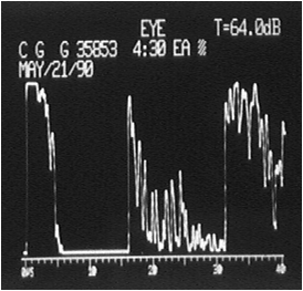
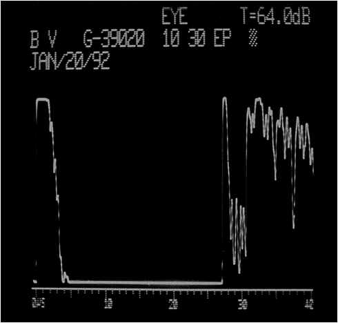
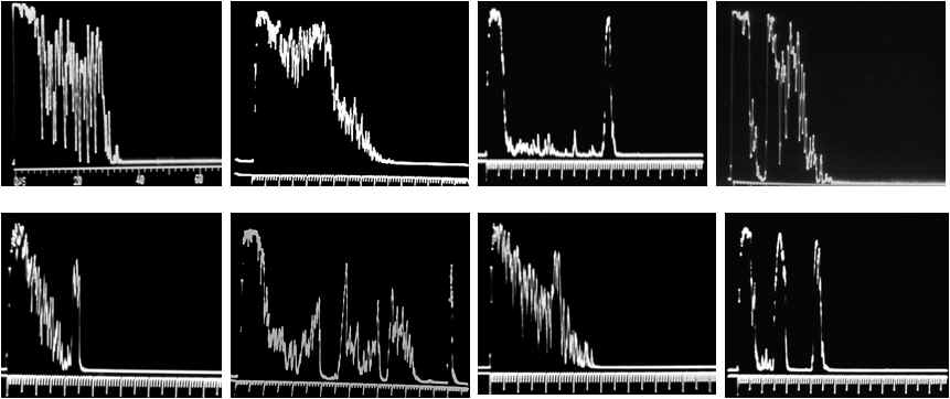
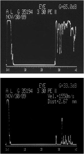
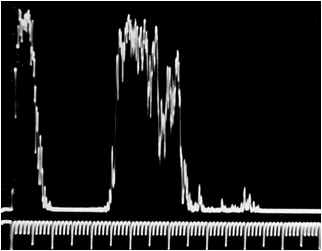
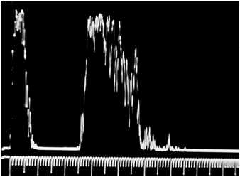
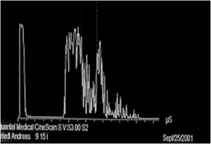
Standardized A-scan: confirms
Standardized
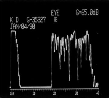
A-scan:
diagnoses
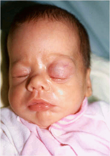
Malignant Melanoma
Malignant
Melanoma
-----------------------------------------------------------------------------------------------------------------------------------------------------------------------------
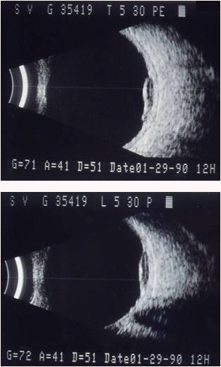
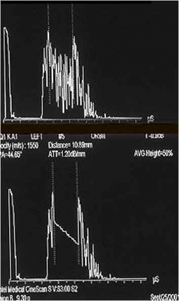
Case # 4
.............................Case # 5..............................
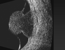
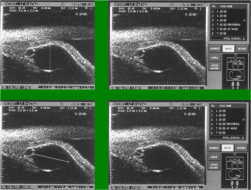
Immersion Method
UBM-B-scan:Solid
Iris Tumor Standardized
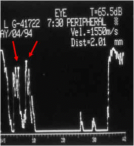
A-scan Diagnosis:
longitudinal scan
malignant
iris melanoma
(between arrows)
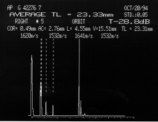
Axial Eye Length Measurement
transverse scan
---------------------------------------------------------------------------------------------------------------------------------------------
other TUMORS of posterior eye segment:
...................................Case # 6...................................
Small
Cell Carcinoma
from the
lung metastatic
to
choroid
OS
-------------------------------------------------------------------------------------------------------------------------------------------------------------------------------
...................................Case # 7...................................
Chorioidal Hemangioma
-------------------------------------------------------------------------------------------------------------------------------------------------------------------------------
...................................Case # 8...................................
Disciform Macula Degeneration
1 1 month follow-up shows marked decrease in height
----------------------------------------------------------------------------------------------------------------------------------------------------------------------------
Examples of ORBITAL TUMORS:
..............................Case # 9...................................
Cavernous Hemangioma (adult type)
---------------------------------------------------------------------------------------------------------------------------------------------------------------------------
...................................Case # 10...................................
Orbital Lymphoma
---------------------------------------------------------------------------------------------------------------------------------------------------------------------------
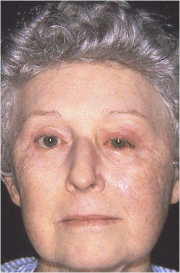
...................................Case # 11..................................
Mixed Cavernous/Capillary Hemangioma in muscle cone of Newborne Baby (Infant-type Hemangioma)
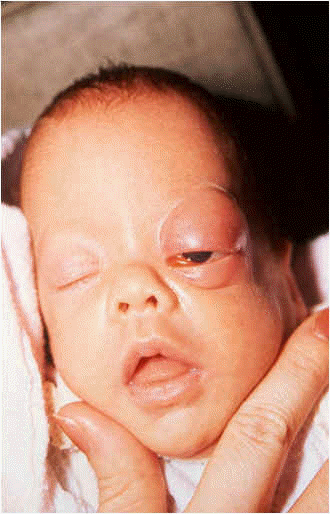
2 days after birth after 1 week of treatment (Prednisone) 5 years later

OS OD OS
------------------------------------------------------------------------------------------------------------------------------------------------------------------------
...................................Case # 12..................................
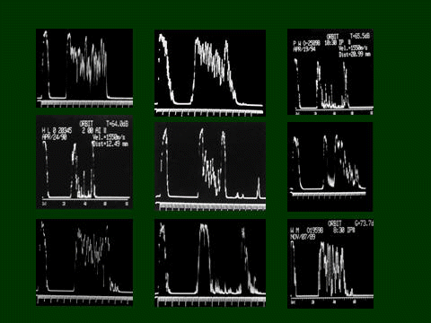
Rhabdomyosarcoma diagnosed with Standardized Echography
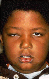
at T+ large angle kappa!
---------------------------------------------------------------------------------------------------------------------------------------------------------------------------
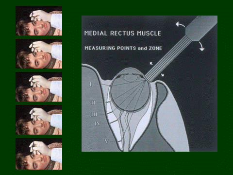
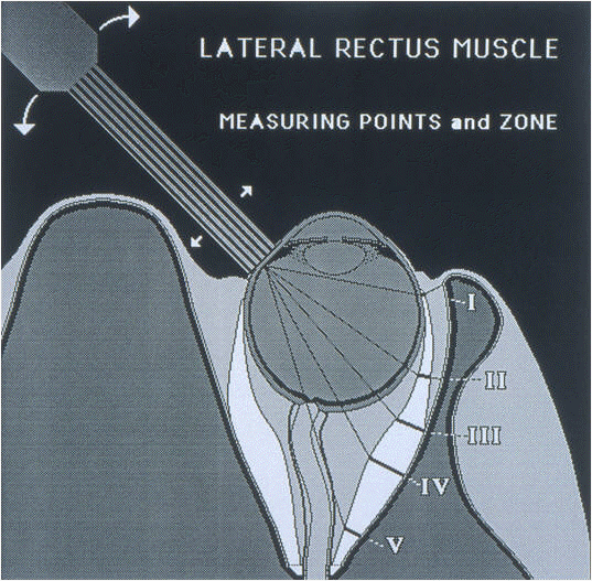
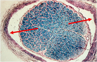
Measuring Points of Extraocular Muscles (example of medial rectus muscle):
---------------------------------------------------------------------------------------------------------------------------------------------------------------------------
...................................Case # 13..................................
bilateral lateral rectus myositis (OD > OS)

---------------------------------------------------------------------------------------------------------------------------------------------------------------------------
...................................Case # 14..................................
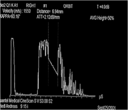
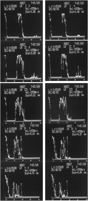
Prostate Cancer (metastasis to left medial rectus muscle)
B-scans (longitudinal) show Tumor within Muscle sheaths A-scans proof Metastatic Carcinoma
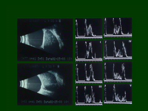
A-scans from around inserting tendon (I) aiming posteriorly over
measuring point III <left column>
over belly region (IV) to orbital apex (V) <right column>
---------------------------------------------------------------------------------------------------------------------------------------------------------------------------
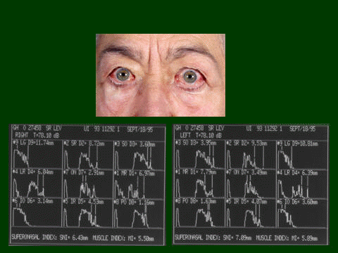
...................................Case # 15..................................
Measurement of e.o. muscles and Grading of the severity of Graves' orbitopathy (orbital profile, muscle index, superonasal index)
---------------------------------------------------------------------------------------------------------------------------------------------------------------------------
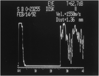
Optic Nerve Measurements:
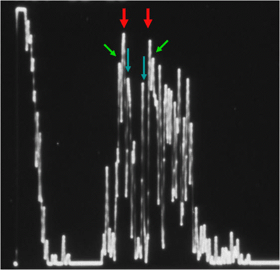
Red Arrows mark Arachnoidal Diameter Blue Arrows mark Pial Diameter Green Arrows mark Dural Diameter
---------------------------------------------------------------------------------------------------------------------------------------------------------------------------
...................................Case # 16..................................
Increased Intracranial Pressure (bilateral optic nerve sheath distensions) due to Intracranial Arachnoidal Cyst
B-scan showing
the increased
Measurement of arachnoidal Measurement of optic disc
elevation unilateral
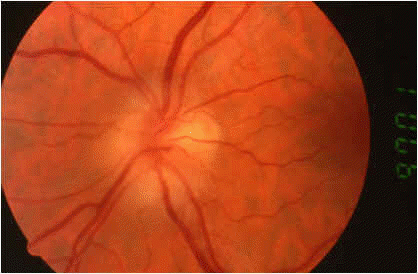
papilledema
subarachnoidal fluid surrounding
diameter (normally < 4.5 mm)
(normally about 0.5
mm)
most anterior optic nerve ("flying bat" sign)
with Standardized A-scan
with Standardized A-scan
Arachnoidal Diameters
Before Drainage of of Cyst Immediately after Drainage 2 months later
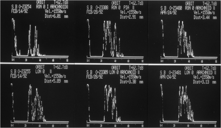
Upper Echograms from Right Optic Nerve Lower Echograms from Left Optic Nerve